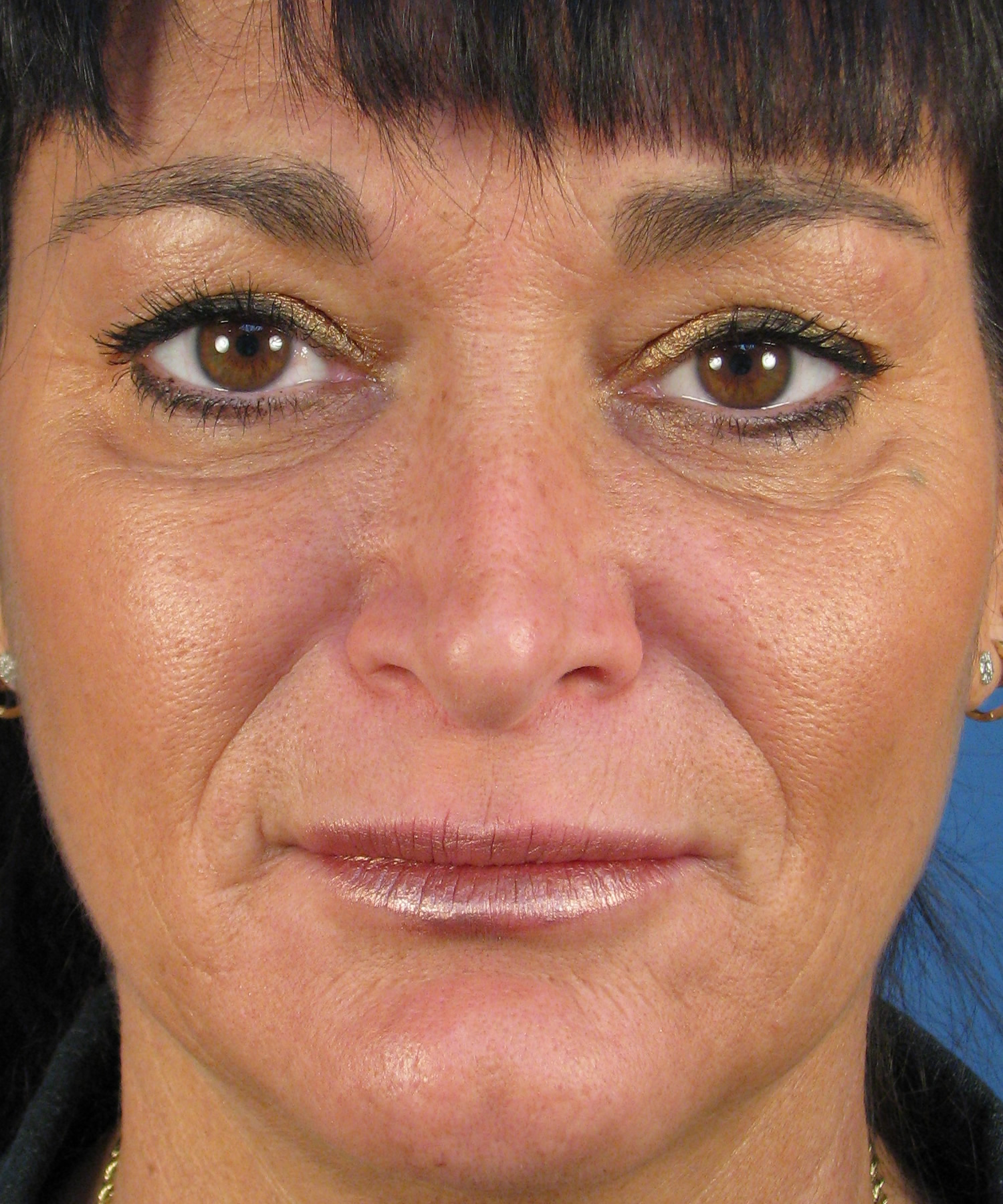
I just finished a revision open rhinoplasty today on this female patient from San Diego. We took some intraoperative photographs to show others what the anatomy looks like when dealing with a pinched nasal tip and hanging columella. This patient had a prior rhinoplasty done by another San Diego plastic surgeon. She was still unhappy with the appearance of her nose and consulted with my office for a revision rhinoplasty.
Preoperative Rhinoplasty Assessment
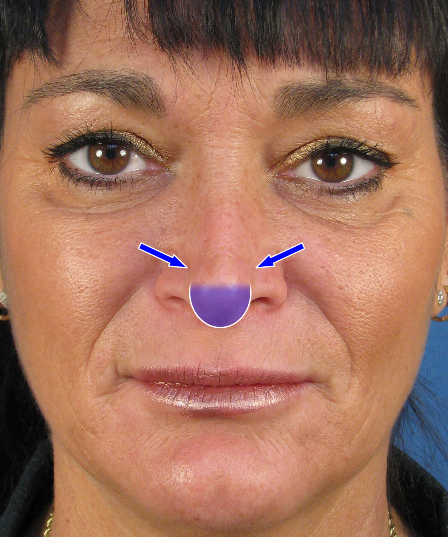
On preoperative rhinoplasty assessment, most of her issues were manifested on the frontal appearance. As you can see below, she had bilateral pinching of the nostrils with her right side being more severely collapsed compared with her left side (see blue arrows). This resulted in an asymmetric tip appearance. In addition, she had what is called a hanging columella. This implies the area in between the nostrils hangs down excessively (see shaded area). These abnormalities resulted from her prior surgery and were readily revealed once we opened up her nose.
![RevRhino75FrontalBefore]()
![RevRhino75FrontalBeforeDiagram]() Intraoperative Photos
Intraoperative Photos
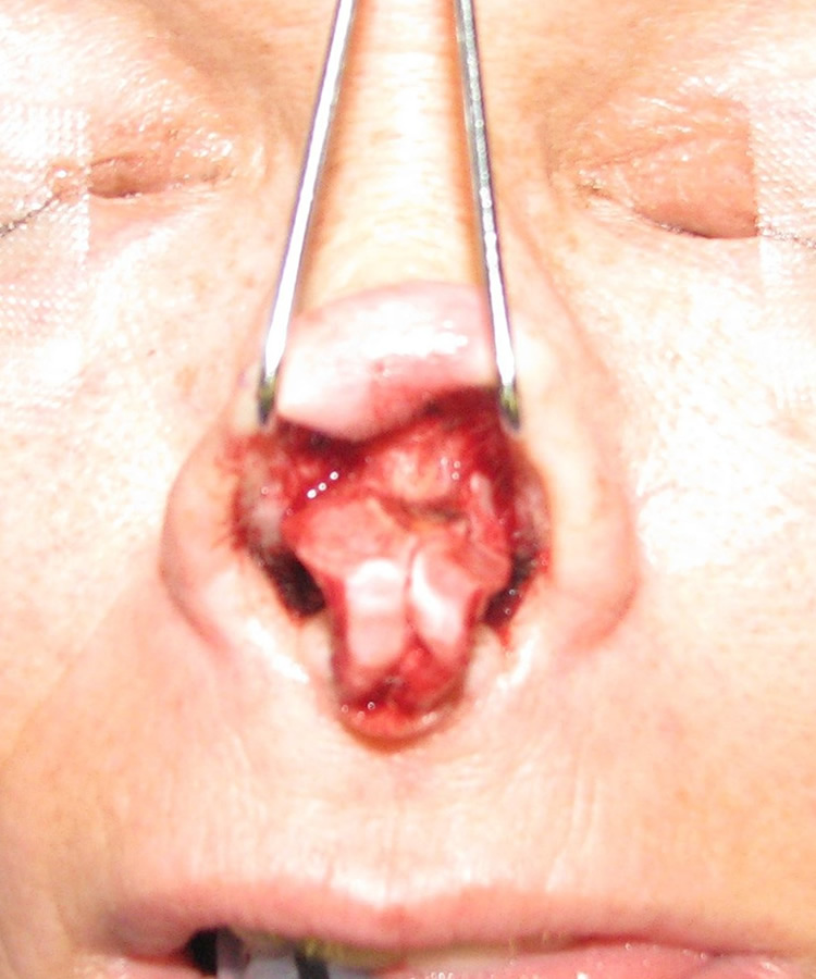
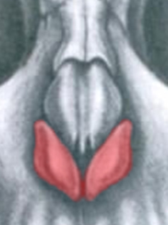 As you will see from her intraoperative rhinoplasty photos, the lower lateral cartilage had been surgically altered clearly explaining the abnormal appearance of her nasal tip. To the left is a quick anatomy reference showing the lower lateral cartilage shaded red. The paired lower lateral cartilages contribute directly to the appearance of the nasal tip. Whenever a plastic surgeon performs a tip rhinoplasty to reshape the tip, the lower lateral cartilage is somehow or another altered. Unfortunately, there are many cases of rhinoplasty where the lower lateral cartilage is surgically altered in a way that compromises the support and position of the tip. For example, in rhinoplasty patients who had too much resected from the lower lateral cartilage, the tip can then pinch inward because the structural support has been excessively compromised. When this occurs, you can see the type of pinching present in this case example as well as development of the hanging columella deformity.
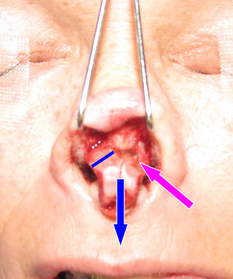
If you look at the first two photos below, you can see that the lower lateral cartilage in this revision rhinoplasty patient of mine has been previously resected, or cut out. In fact, the pink arrow points to her left lower lateral cartilage, which had been resected more aggressively and was trimmed irregularly versus her right side. The blue line spanning the lower lateral cartilage shows what is remaining from her original lower lateral cartilage. By actual measurement, this was about 6 mm wide. Unfortunately, when a rhinoplasty surgeon leaves only this remaining width of cartilage, it is insufficient to maintain proper shape and position. In this case, the narrow remaining segment of cartilage scarred upward at the same time it pinched inward. The medial, or center, segment of the lower lateral cartilage (called the medial crus) had been altered such that it was no longer connected to the nasal septum. In the setting of excess cartilage resection, this latter issue resulted in the hanging columella deformity. You can see from the blue arrow that the cartilage almost appears too heavy as if it were hanging down off the rest of the nasal tip.
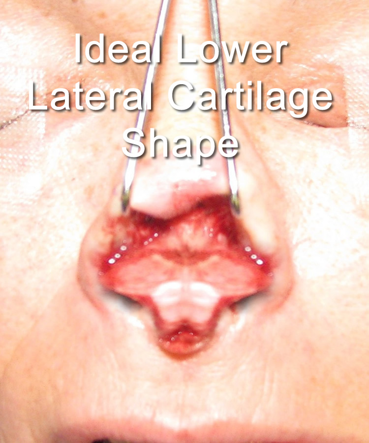
In the third photo below, I have morphed her intraoperative rhinoplasty photo to show more what her nasal tip cartilage should look like ideally. The width of the lower lateral cartilage should be nearly twice as wide as shown. With this amount of cartilage, there is sufficient support for the nasal tip so that pinching is not a real concern. In addition, the medial crus should be shorter so that the columella does not look like its hanging. In this particular case, I fully reconstructed her nasal tip using an open revision rhinoplasty technique with ear cartilage grafting. Fortunately, we were able to achieve a very nice surgical result. I look forward to showing you the postoperative photos 10-12 months from now. I hope this blog entry was helpful in showing you potential revision rhinoplasty patients what a pinched nasal tip and hanging columella deformity looks like in surgery!

