Facial Plastic Surgery
Functional upper blepharoplasty surgery refers to an eyelid reshaping procedure to help improve poor vision due primarily to excess upper eyelid skin.
- How An Upper Eyelid Lift Can Improve Vision
- Can An Upper Eyelid Surgery Improve My Appearance?
- Functional Upper Eyelid Blepharoplasty Procedure
- Insurance Coverage For An Upper Eyelid Lift
- Typical Functional Upper Eyelid Lift Patient
- Photo Results of Improved Vision After Lift
- Am I A Candidate For An Upper Eyelid Lift?
How An Upper Eyelid Lift Can Improve Vision
The following photos demonstrate more graphically what an upper eyelid lift can do for people who have impaired peripheral vision from excess upper eyelid skin. The top photo shows the effect of having excess, weighted skin hanging down along the upper eyelid. As you can appreciate, the black shadowing corresponds to how much of the peripheral vision can be blocked in some patients because of the draping effect. The bottom photo demonstrates the type of improvement that is desired after undergoing an upper eyelid lift to remove the excess, heavy skin.

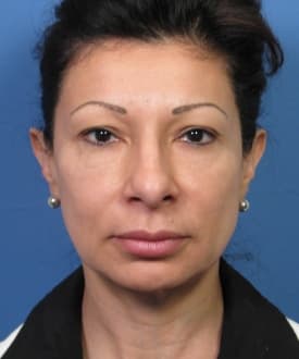
Before Upper Blepharoplasty

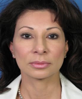
After Upper Blepharoplasty

As shown in the adjacent photo diagram (A), the lid margin (red arrow) is typically positioned 1-2 mm into the iris. Another way to measure the upper eyelid position is by what is called the marginal reflex distance, or MRD. This measurement is taken between the light reflex seen just to the sides of the pupil and the upper eyelid margin. Normally, the MRD is approximately 4 mm with decreasing numbers representing more ptosis of the upper eyelid. On the right side (B) of the photo diagram, the upper eyelid has been modified digitally to show what a ptotic upper eyelid would look like in comparison. As you can see, the eyelid margin has now fallen well into the colored portion of the eye and is even quite close to being into the pupil. This type of abnormality can obviously contribute to impaired vision. And in cases where dermatochalasia also exists, ptosis can significantly worsen the problem.
Ptosis of the upper eyelid is not due to excess weighted skin. In most facial plastic surgery practices, it is more commonly due to an abnormality with a small muscle within the upper eyelid (called the levator muscle) and a connective tissue attached to the muscle (called the levator aponeurosis). Therefore, the corrective procedure to fix ptosis of the upper eyelid includes some type of surgery involving the levator muscle and aponeurosis. This is usually in the form of a tightening procedure that is performed often times through the natural upper eyelid crease. In cases where dermatochalasia is present, both procedures can be done during one operation – removal of the excess upper eyelid skin combined with ptosis repair eyelid surgery.
Can Functional Upper Eyelid Surgery Improve My Appearance?
Many patients who are considering upper eyelid surgery to improve their vision often ask whether or not they are going to look better from a cosmetic standpoint. Fortunately, one of the byproducts of having functional upper eyelid surgery is that there is likely going to be a passive cosmetic enhancement to the eyes. Since the procedure is done in a very similar manner to esthetic upper eyelid blepharoplasty, there is often times a significant cosmetic improvement that is seen.
Functional Upper Eyelid Blepharoplasty
A blepharoplasty procedure to help improve the visual field is done in a similar fashion as is done for cosmetic enhancement of the upper eyelid. The surgery can usually be done with either local anesthesia alone or a light sedative. Either way, this is done on an outpatient basis where the patient can go home shortly after the eyelid lift has been completed. The upper eyelid is lifted by making an incision based on the natural eyelid crease so that the final scar is least visible as possible. The excess upper eyelid skin is then removed while leaving behind a sufficient amount to allow proper closure of the eye. By removing the excessive amount of skin that is bunched up along the eyelid, the shading effect can be reversed and the peripheral vision can be greatly improved.
Ptosis repair can also be performed through an upper eyelid incision and can be done at the same time as removal of the excess upper eyelid skin. Ptosis repair requires going deeper into the upper eyelid soft tissue to expose the levator muscle and aponeurosis. The aponeurosis is typically shortened, or tightened, as part of the repair process. This is accomplished by strategically placing very fine sutures into the levator aponeurosis. In doing so, the upper eyelid is lifted to a more appropriate level thereby improving the opening of the eye.
Sutures, or surgical stitches, are used to close the incision. These are typically removed in 5-6 days. Some upper eyelid lift patients notice a mild to moderate amount of swelling during the first 72 hours of healing, but appear fairly presentable in public by day 7-10.
Insurance Coverage For An Upper Eyelid Lift
Many patients seeking blepharoplasty surgery inquire whether or not their health insurance can be used to provide coverage to reshape their upper eyelids. The answer is that some insurance companies will, indeed, pay to have an upper eyelid lift to help improve your vision. In essentially all of these cases, there has to be specific medical documentation that confirms your upper eyelids as the source of the visual problem before it is medially indicated to undergo an upper blepharoplasty. Health insurance companies typically demand a ‘visual field test’ to obtain more objective evidence that the upper eyelids are impairing your vision. A visual field test is usually done for this purpose with the eyes untaped then taped. Basically, the visual field testing involves the patient looking into a box with one eye covered. A series of brief light flashes are presented to the patient in the distribution of the normal visual field. The patient is asked to confirm whether or not they saw the flash by pressing a button. If the patient fails to press the button when the flash of light is presented it is assumed they did not see it in their visual field. The patient’s excess upper eyelid skin is then taped upward to simulate what an upper eyelid lift might accomplish. The testing is then repeated. If there is a significant improvement noted in the number of light flashes seen with the eyelid taped up, the test is considered positive – confirming the potential benefit of undergoing a functional upper eyelid lift, or functional blepharoplasty.
Typical Upper Eyelid Lift Patient
 The typical patient who presents for an upper eyelid lift to improve their vision has a moderate to severe amount of excess upper eyelid skin. Many patients refer to this upper eyelid skin as being redundant or having too many folds. Others refer to this as eyelid hooding. In medical terms this is referred to as dermatochalasis or dermatochalasia, which refers to excessive amount of eyelid skin. This type of redundant upper eyelid skin can result or be aggravated from a variety of factors, which include sun exposure, allergies, constant rubbing, and congenital predisposition to sagging skin.
The adjacent patient photo is a good example of upper eyelid skin redundancy that is contributing to blocked vision. As you can see by the arrows, this male blepharoplasty patient from San Diego has an excess amount of skin overlying his upper eyelids on both sides. In fact, in his case you can barely even see the natural upper eyelid crease. The blue dotted line indicates where the upper eyelid crease normally exists. As you can see, there is such an excess of upper eyelid skin in this patient that the natural crease has been nearly completely obscured. If you will also notice, this patient’s eyelashes are also covered by the excess skin folds hanging down over the upper eyelid. With this type of situation, it is not surprising that this patient has trouble with his peripheral vision due to the skin folds hanging over the upper eyelid.
Photo Results of Improved Vision After Lift
The following photo examples demonstrate the type of improvement that can potentially result from an upper eyelid lift to address problems with peripheral vision. These upper eyelid blepharoplasty photos represent actual patients of The Hilinski Clinic of Facial Plastic Surgery in San Diego, CA.


Am I A Candidate For A Functional Upper Eyelid Lift?
If you are considering an upper eyelid lift to help with impaired vision, contact our San Diego plastic surgery office today. Dr. Hilinski currently accepts a variety of PPO health insurance plans for medically indicated upper eyelid surgery. This includes, but is not limited to, Blue Cross, Blue Shield, United Healthcare, Aetna Healthcare and Tricare insurance plans.
Planning Your Functional Upper Eyelid Surgery

Average Cost: $5,500 – $6,500
Average Procedure Time: 1 – 2 hours
Average Recovery Time: 7 – 10 days
Post-Op Consultation: 3 months
*Procedure pricing and results with Dr. Hilinski may vary. Your personalized treatment plan and pricing will be determined at your consultation appointment.
Dr. John Hilinski has either authored or reviewed and approved this content.