As a leading expert in nose surgery and facial reconstruction, Dr. John Hilinski specializes in both cosmetic and reconstructive nose surgery following skin cancer removal. Many patients come to him for his vast experience in rhinoplasty and revision rhinoplasty, making him the go-to surgeon for those needing reconstructive nose surgery. This case showcases how Dr. Hilinski’s expertise helps patients regain their appearance and function following skin cancer treatment.
Dr. Hilinski, Nicole, and team are absolutely amazing! Dr. Hilinski is very knowledgeable, takes the time to thoroughly explain procedures, and has gone above and beyond with patient care during my procedure and throughout all of my follow-ups.
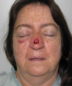
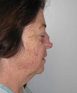
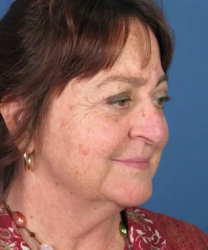
Case Study: Skin Cancer Reconstruction After MOHS Surgery
In this case, a patient from San Diego, CA, was diagnosed with skin cancer on the bridge of her nose. After her dermatologist performed MOHS surgery to remove the cancer, she was referred to Dr. Hilinski for reconstructive surgery. MOHS surgery is a common technique used to remove cancer while preserving as much healthy tissue as possible. However, in this patient’s case, the procedure left a significant defect in the middle of her nasal bridge. The defect involved both the skin and the underlying bone and cartilage.


Faced with concerns about both function and appearance, the patient sought Dr. Hilinski’s expertise. His specialized training and experience in reconstructive rhinoplasty allowed him to develop a successful surgical plan to restore her nose’s structure and aesthetics.
Paramedian Forehead Flap Surgery
Given the complexity and size of the defect, Dr. Hilinski chose to perform a paramedian forehead flap. This advanced reconstructive technique involves transferring a section of skin, along with its blood supply, from the forehead to the nose. This method provides the nose with robust tissue and a reliable blood supply, ensuring a successful outcome.
Skin grafts were not an option due to the depth of the defect and the need for cartilage and bone reconstruction. With the paramedian forehead flap, Dr. Hilinski was able to effectively rebuild the nose with a natural look while preserving functionality.
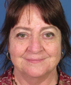
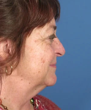
Before and After Photos
The patient’s journey included multiple stages of surgery, allowing her nose to heal and integrate the flap. As shown in her before and after photos, Dr. Hilinski’s technique restored both the aesthetic appearance of her nose and the underlying structure. Today, she has no visible signs of the cancer removal surgery.


FAQs: Skin Cancer Reconstruction
Skin cancer reconstruction involves surgical techniques to repair the face after skin cancer removal. This can involve tissue grafts, flaps, and structural repair to restore both function and appearance.
Anyone who has undergone skin cancer removal and is left with a significant facial defect, particularly in the nose, may benefit from reconstructive surgery.
A paramedian forehead flap is a surgical technique where tissue from the forehead, along with its blood supply, is transferred to another area, such as the nose, to repair defects after skin cancer removal.
Recovery after a forehead flap surgery can take several weeks. The initial stage involves allowing the flap to integrate into its new location, followed by a second stage where the flap is refined for optimal results.
In most cases, insurance will cover reconstructive surgery following skin cancer removal. It is recommended to check with your provider to confirm coverage.
While scars are inevitable, Dr. Hilinski uses advanced techniques to minimize their appearance and blend them with the natural contours of your face.
Yes, in some cases, reconstructive surgery can be combined with cosmetic procedures to enhance the overall outcome and achieve a more harmonious facial appearance.

Why Choose Dr. John Hilinski for Skin Cancer Reconstruction?
Dr. John Hilinski is a board-certified facial plastic surgeon with dual certifications in Facial Plastic and Reconstructive Surgery and Head and Neck Surgery. His extensive training and years of experience make him uniquely qualified to handle complex cases of facial reconstruction, particularly in sensitive areas like the nose. Dr. Hilinski has earned a reputation as one of San Diego’s top facial plastic surgeons for both cosmetic and reconstructive procedures. His advanced surgical skills and patient-focused approach ensure the best possible outcomes for each individual.
Schedule a Consultation for Skin Cancer Reconstruction
If you’ve undergone MOHS surgery or another skin cancer treatment and are seeking reconstructive surgery, schedule a consultation with Dr. John Hilinski in San Diego. His experience in both cosmetic and reconstructive nose surgery will help you restore your facial appearance and confidence.