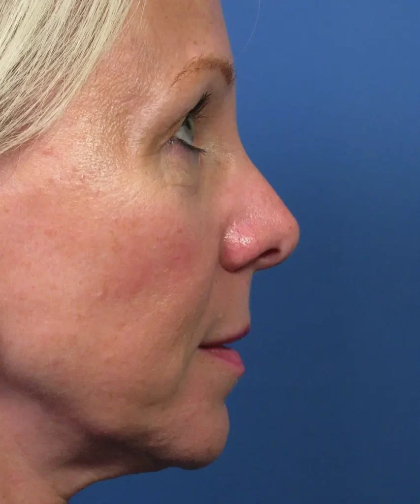
This is a case example of a patient who desired to have her nose reshaped following two prior unsuccessful attempts performed by a different plastic surgeon. Her surgeries were performed over 10 years prior. Since then, her nose developed asymmetric collapse of the tip region – with more collapse seen on her right side. This was bothering her enough that she wanted to have the tip reshaped to be more symmetrical. In addition, she desired to have the bridge brought down just a bit.
Rhinoplasty Exam
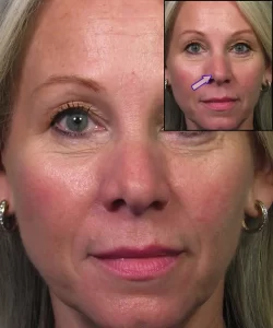
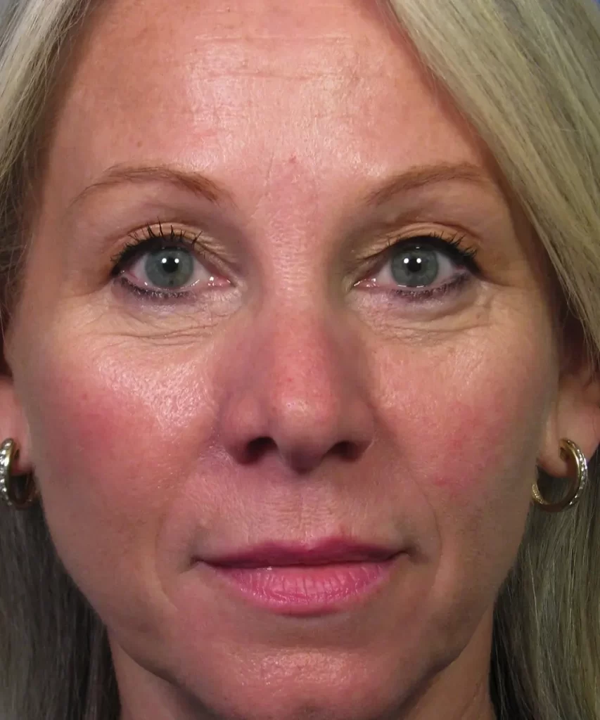
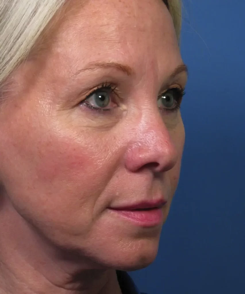
Her preoperative examination demonstrated an asymmetric nasal tip with more collapse seen on her right side. You can see this best on her frontal view as more shadowing through the right tip region. In the adjacent photo diagram, this is indicated by the arrow pointing to the specific area of collapse. This indentation results in more shadowing on her right side – an area commonly referred to as the soft triangle of the nose. In addition, this type of collapse can negatively impact nasal function since the nostril rim is also compromised in terms of structural integrity. The patient and I discussed in detail what might be contributing to this type of nasal tip deformity following prior rhinoplasty. We also reviewed my recommendations for a revision rhinoplasty procedure to include an open approach (where the skin is reflected upwards) for optimal exposure. In addition, she would likely need cartilage grafting to help restore proper structural support of the nasal tip.
Rhinoplasty Surgical Findings
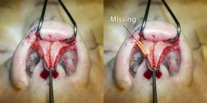
These photos provided here show what I saw after opening her nose up during the actual surgery. This photo, taken from underneath the nose, highlights the lower lateral cartilages, which largely dictate what the tip of the nose looks like in real life. As you can see here, there is a difference between the right and left sides where her left lower lateral cartilage is wider than her right one. This is because the prior surgeon had resected, or cut out, more cartilage on the one side – leaving her nasal tip with two different lower lateral cartilages. The yellow outlined polygon shows approximately what is missing from her right lower lateral cartilage when compared to the opposite side. In some rhinoplasty patients, such a difference between the two sides may not be significant. But in this case, the relative lack of cartilage on the one side translated into a notable clinical difference in terms of shape and contour.
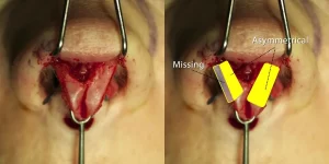
When you look at the same two lower lateral cartilages from above, you can more readily appreciate the differences just pointed out. As you can see in the adjacent photo diagram, her right side is much thinner than her left side. The relative difference is indicated by the yellow outlined polygon, which, again, is a significant portion of cartilage to be missing from her tip region. Now that you can visualize it (as I did in surgery), you gain a better understanding of how asymmetrical her two lower lateral cartilages are in size and shape.
Rhinoplasty Changes Made
So what did I do to help reshape her nasal tip during the rhinoplasty? First, I strategically placed several permanent sutures into the tip cartilage to help make the overall shape of the nasal tip more defined. These are technically called ‘dome binding sutures’ in rhinoplasty parlance. This is represented in the adjacent photo by the blue threads that can be seen sticking out of the cartilage. The darker purple indicates the surgical markings that were made to help guide placement of the sutures. If you compare the overall shape of the lower lateral cartilages to what they looked like before reshaping, you can readily appreciate the change in conformation. Meaning, the cartilage is now less rounded and more angulated. This was done to give her nasal tip a more refined shape.
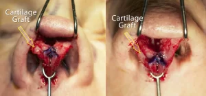
But what about the missing cartilage segment noted previously? In order to address this, I ended up performing a septoplasty to get some cartilage that could be used to help reconstruct the missing segment of the lower lateral cartilage. This septal cartilage was repurposed to restore proper support of the lower lateral cartilage. As is shown in the adjacent photo diagram (by the yellow outlined arrows), cartilage grafting was performed. The septal cartilage was sewn into the area where there was missing cartilage to help provide a more symmetrical framework to the nasal tip. Combining the dome binding sutures with septal cartilage grafting, I was able to completely reconstruct her nasal tip.
Beyond the nasal tip changes made, I also performed reduction of her nasal bridge height along with narrowing of her nasal bones.
Rhinoplasty Before and After
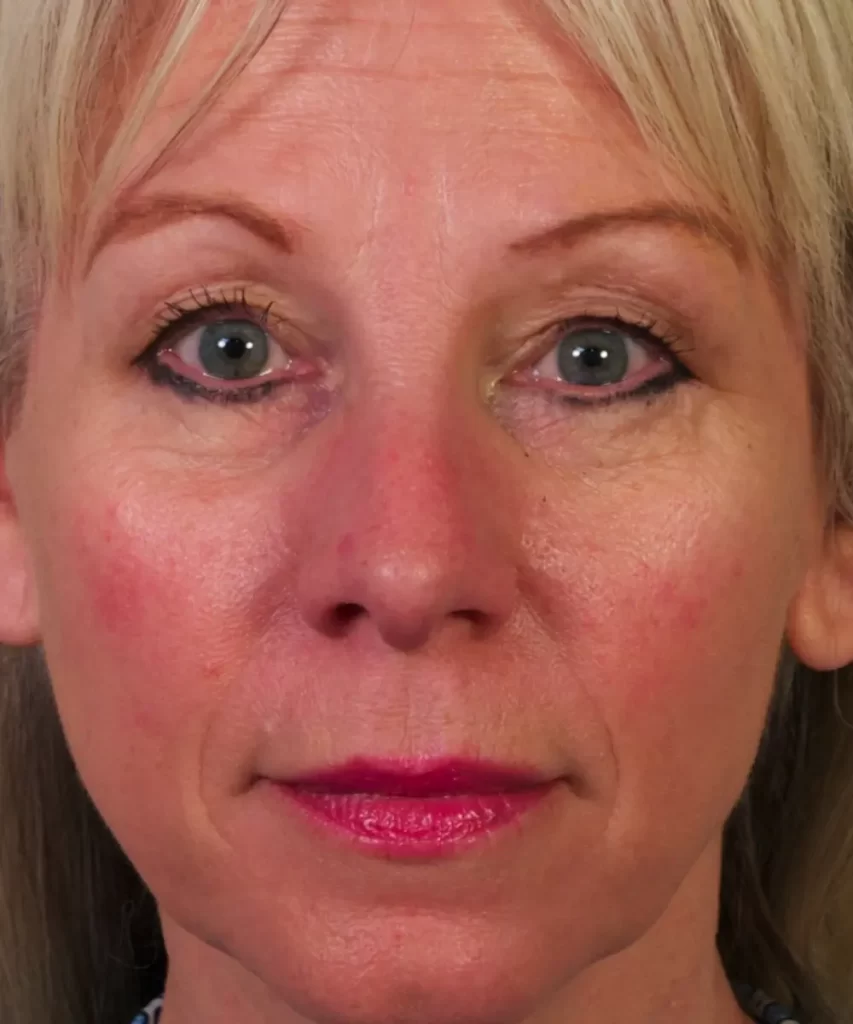
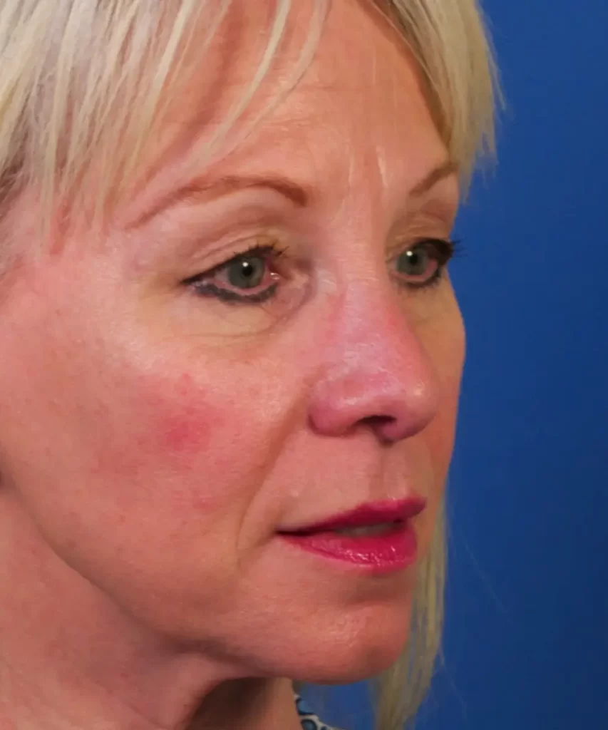
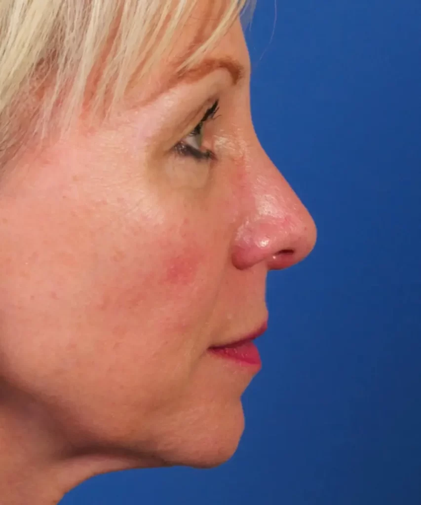
Here are her rhinoplasty before and after photos that show the overall visual change. As you can see, she no longer has the abnormal shadowing through her soft triangle region, which was seen prior to surgery. Her nasal tip is now more symmetrical with proper support and structure on both sides. In addition, her nasal tip now is more defined and refined. Of utmost importance, her nose has been improved while still looking quite natural for her face.