In some cases of cosmetic rhinoplasty, the changes made to the shape of the nose can have a much more dramatic impact on the overall facial appearance. This is a great example of such a case.
This young lady from San Diego consulted with me in the hopes of having cosmetic nose reshaping surgery. More specifically, she did not like the bump on the bridge and the wide appearance as seen from the frontal view. In addition, she desired tip reshaping to match the changes being made to the nasal bridge.
We discussed at length the various changes that were possible in terms of cosmetic rhinoplasty surgery. This included the goal of trying to reduce the bump, narrow the bridge, and define the tip – all the while creating a nose that looked in natural balance with her underlying facial features.
Rhinoplasty Computer Imaging
As part of the rhinoplasty consultation process, the patient was provided computer imaging of what her nose may look like following surgery. These images are shown here. On her frontal view, the idea was to narrow the nasal bones to create more shadowing along the sides of the nose – thus giving her more definition as seen from the front.


Now many patients may look at the computer enhanced photo from the front and wonder why there is not a more dramatic change. There are number of reasons to explain this. One of them relates to the inherent limitations of computer imaging. Making computer enhanced modifications to the nose from the front view can be fairly difficult to do in some cases. The program certainly allows me to create additional shadows while erasing others. But doing this in a manner where the visual result is realistic is the challenge. Imaging the lateral view is much easier given the contrasting background color. Unfortunately, all too often you see other rhinoplasty surgeons perform imaging of someone’s nose where the computer enhanced version is simply unrealistic. In fact, in some offices the surgeon is not even the one doing the imaging for the patient. If rhinoplasty surgery is so complex and individualized, how can someone with absolutely no surgical training be the person showing you what might happen with a complex series of surgical maneuvers? Making sure the computer enhanced images are realistic and achievable is really important when it ultimately comes to assessing the surgical outcome of a particular case.


Therefore, I am a big proponent of trying to ‘under image’ rather than ‘over image’ patients’ photos. Meaning, I would rather show a rhinoplasty patient a more conservative computer enhanced image that they would be satisfied with after cosmetic nose surgery. If I can provide them an even better result than the computer image, then great. But I would want the patient going into surgery with the mindset they will still be content with the more conservative result as imaged with the computer program.
With the above imaged photos presented to her, this patient was quite happy with the surgical goals I had outlined and was ready to proceed with her rhinoplasty surgery.
Rhinoplasty Hump Reduction
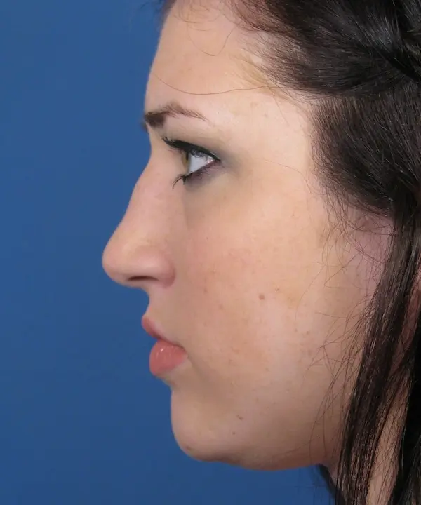
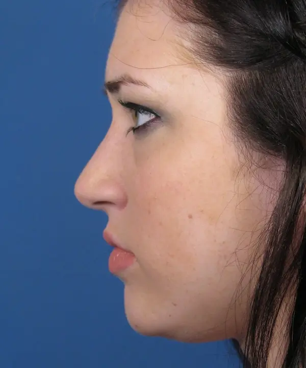
One of the main goals in this particular case was to reduce the dorsal hump – or bump along the upper bridge. As was shown to her in the imaged photos, the goal was to create a straighter looking profile view without over-reducing her bridge, thus avoiding a scooped appearance. This was done during her surgery using a rasping technique whereby the excess bony prominence is sanded down. Once this was done, the nasal bones were then fractured, or intentionally broken, to narrow the bridge. If the nasal bones were not broken in her case, the nose would appear much wider from the front since the bridge height was taken down.
This part of her rhinoplasty procedures brings up a great point that is worth further discussion. Unfortunately, many patients (and even plastic surgeons) are under the false impression that hump reduction rhinoplasty, or removal of a hump from the nose, is a ‘simple thing’ to perform. In fact, I hear it all the time from my patients that other surgeons comment to them regarding how easy it is to ‘just take down the bump’ on the bridge of their nose. Sadly, it is this type of erroneous thinking that is what gets many patients (and plastic surgeons) into trouble. The cold, hard truth is that hump reduction in rhinoplasty is one of the most challenging types of cosmetic nose surgery to perform while achieving consistently, beautiful results.
The reasoning behind this relates to the fact that the bump, or nasal hump, is not just an issue that impacts the profile view of a rhinoplasty patient. Almost any nasal hump impacts both the side view as well as the front view of any patient. Once the nasal hump has been removed or sanded down, you also have to take into consideration what this does to the front view of that patient. In a good majority of cases, once the hump has been reduced, the surgeon will then break the bones in order to maintain an appropriate width to the nose as seen from the front. When you begin breaking the bones, this triggers a whole new set of variables involved in the surgery and healing process that can significantly impact how the nose looks from the frontal view. For example, breaking the bones in this situation requires cutting the nasal pyramid bone on both sides. As the bones are repositioned it should be done in a manner where the bridge alignment is kept as symmetrical as possible. If the bones start off relatively symmetrical, the alignment needs to be maintained. If the bones start off crooked, they should be moved such that the alignment is made more symmetrical. To complicate matters further, the lower part of the nose will usually follow where the upper part moves in terms of alignment. Meaning, shifting the upper one-third of the nose (the bones) will often times impact the position and shape of the lower two-thirds of the nose. So, as you can now appreciate, there is somewhat of a domino effect in play when a hump is removed – the bones then need to be broken, which can affect the alignment of the bridge, which can then alter the shape of the middle and lower part of the nose. This series of considerations hardly makes hump removal a simple proposition.
Proof of this concept can be seen in the many plastic surgery and rhinoplasty galleries online that show before and after photos of a patient undergoing hump reduction surgery. In a great number of these cases, the plastic surgeon will only show you the profile, or side, view of the nose before and after hump reshaping. But, as a potential rhinoplasty patient, you should really ask yourself, “Why did the surgeon not show the front view of that same patient?” If the frontal view of that patient looked improved (or even the same), you would think the surgeon would be showing you that view as well, right? The reason it is not online on their website is usually because the front view doesn’t look so good following hump reduction rhinoplasty. That’s because it is, indeed, quite a challenge getting the frontal view of the nose to look symmetrical and proportional following hump reduction. I have gone so far to even dare potential rhinoplasty patients to call those surgeons’ offices and inquire about seeing the frontal view of those same patients on their websites. In all likelihood, the frontal views of those hump reduction patients will not be shown to you because they simply don’t look right. If you look at my gallery of rhinoplasty photo examples, you will see the patients undergoing hump reduction have a frontal view accompanying the profile view in nearly all cases. That is because having multiple photo angles of the rhinoplasty patient undergoing reduction of the hump deformity tells a much more accurate story of what type of changes were really made to that particular nose.
Rhinoplasty Transformation
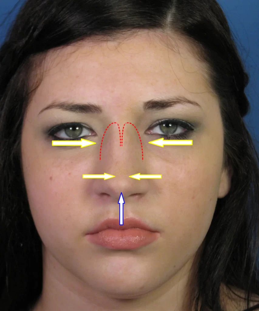
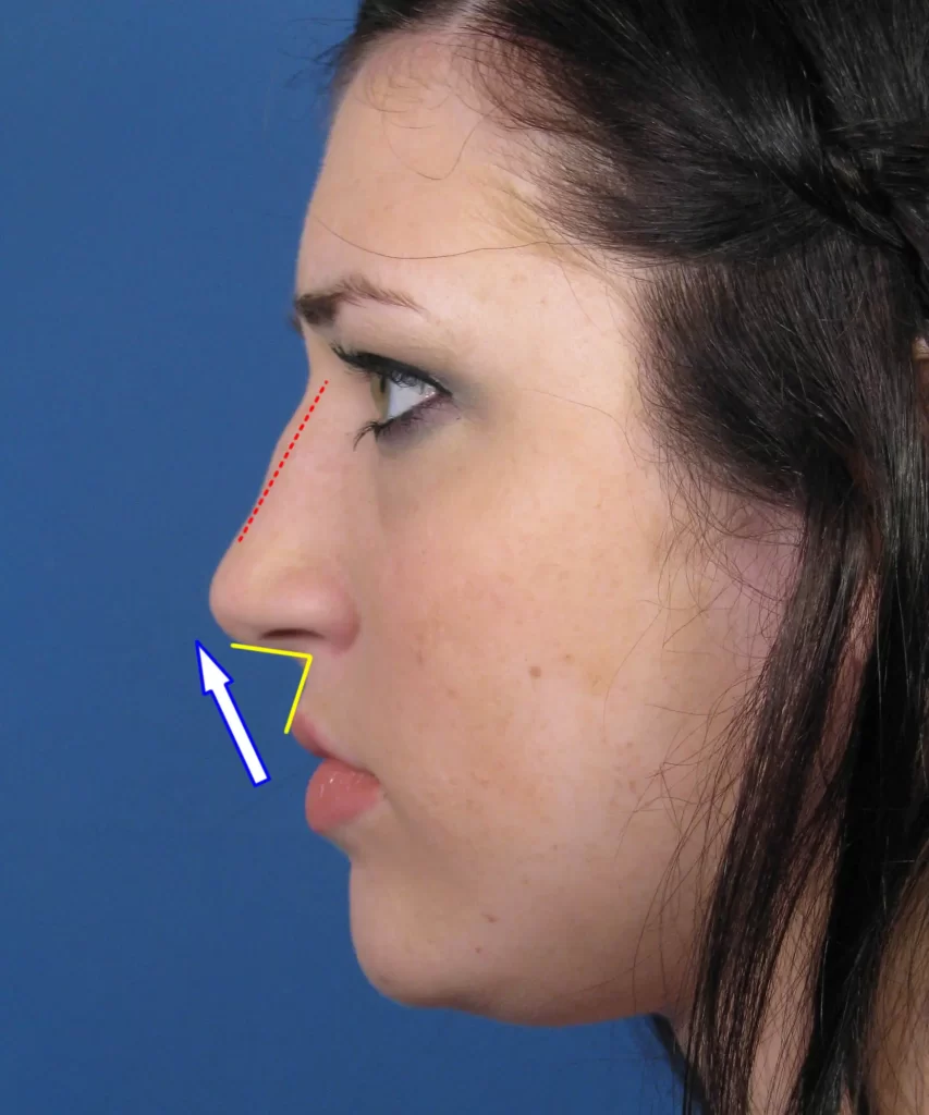
In this rhinoplasty patient, the hump reduction was done in conjunction with tip reshaping as was planned. The amount of hump reduction of the bridge can be seen diagrammatically in the adjacent photo diagram as designated by the red dashed line on her profile view. As was discussed previously, her nasal bones then required osteotomies (intentional cutting) as designated by the red dashed curves seen on her frontal view). The nasal bones were then pushed inward as shown by the yellow arrows to create improved shadowing of the upper bridge region. The nasal tip was refined primarily by use of permanent sutures that were strategically placed to reshape her native cartilage. At the same time, I brought her nasal tip upward as shown by the blue arrows. If you look at her profile view, you can see that her nasolabial angle (the angle formed by the junction of the bottom of the nose and the upper lip) is just under 90 degrees. Ideally, in a female this nasolabial angle should be closer to 95-105 degrees. Bringing her nasal tip up just a bit would open this angle up, thus creating a more pleasing appearance. All of these changes were done using an open rhinoplasty approach that provided me with optimal exposure of her nasal framework.
Cosmetic Rhinoplasty Results
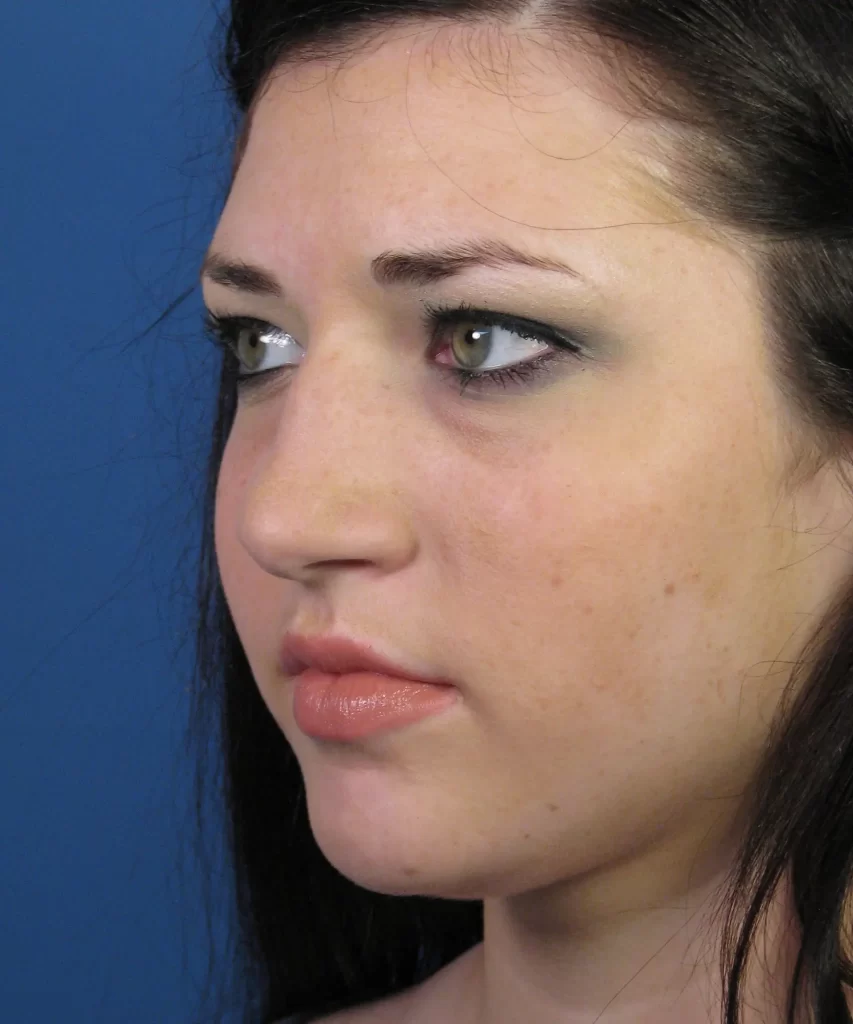
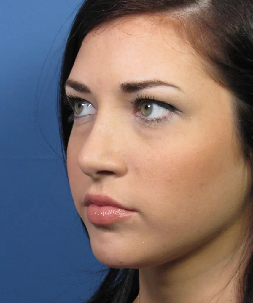
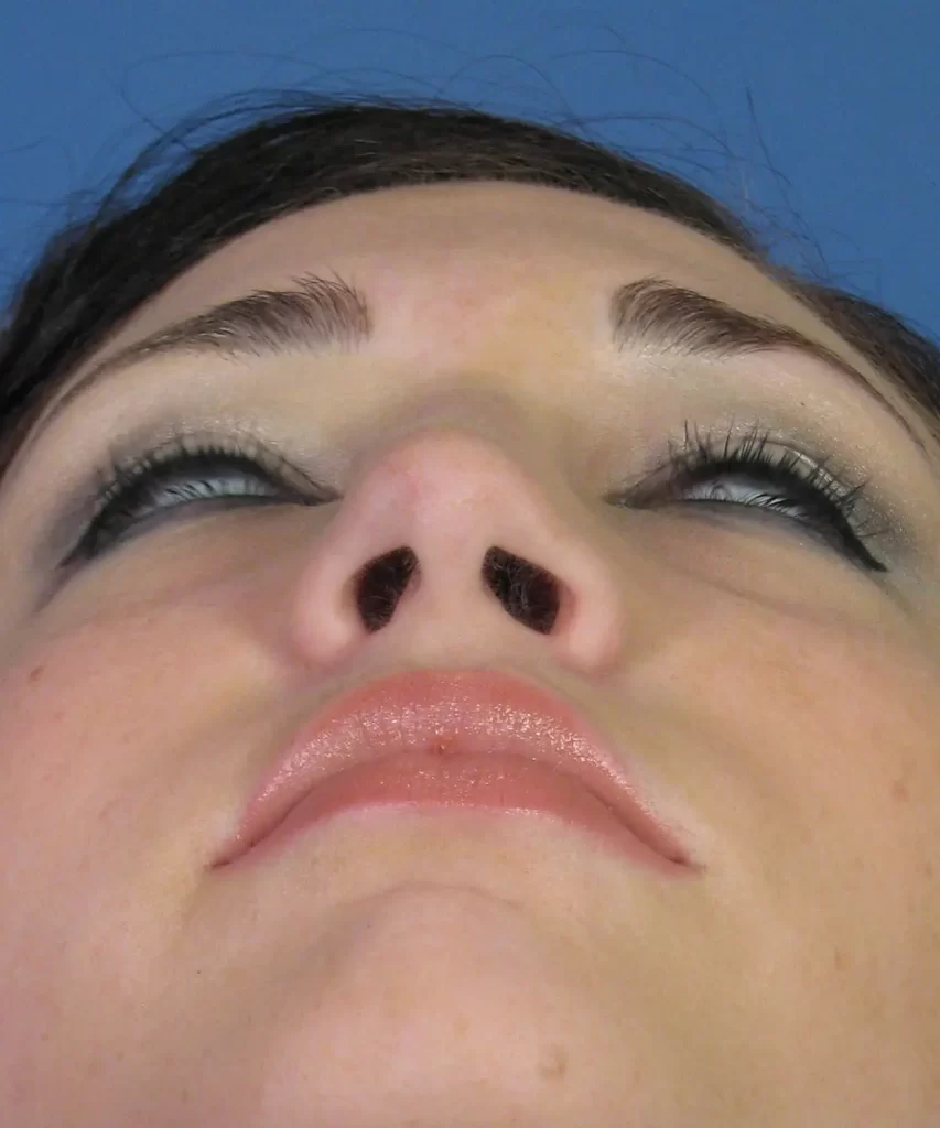
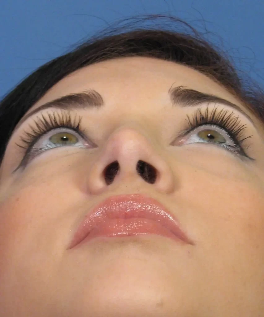
Shown here are the rhinoplasty results following the surgical changes noted above. As you can see, her nose has been transformed to create a fairly obvious change in her facial appearance. On the front view, her bridge and tip are now more narrow with improved shadowing on both sides due to increased definition. In addition, her nasal tip has been brought upward just enough to match the other changes noted. Her oblique (45 degree) view really shows how much the bridge has been brought down as the previously noted bump is now gone. Her profile view confirms this transformation with a bridge that is now much straighter and a tip that is more ideally positioned as seen from the side. Her base view (under the nose) shows how the overall shape of her nasal tip has been transformed. Previously her base view showed a more rounded, wide shape to the tip region. Following cosmetic rhinoplasty, it is now more ideally like a soft triangle.


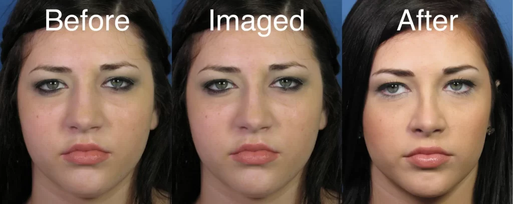
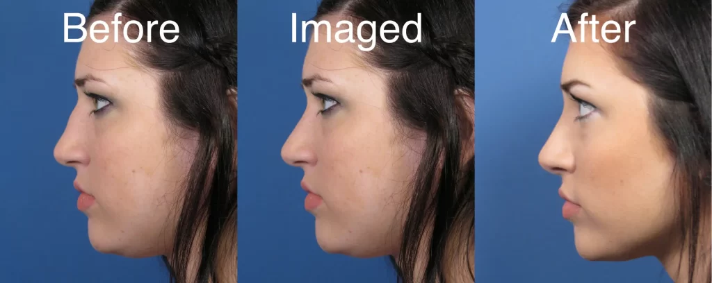
Rhinoplasty Imaged Photos
We previously discussed the exercise of imaging rhinoplasty patients’ photos to give them an idea of what they might expect following surgery. So here are the imaged photos of this particular rhinoplasty patient shown in sequence with her preop photo and actual surgical result. As you can see on both the frontal and profile views, her final rhinoplasty result is fairly close to what was envisioned during the computer imaging process. In fact, as noted above, I somewhat ‘under imaged’ her photos preoperatively. In the end, though, I believe the rhinoplasty surgery provided her with a nose that was even better than the imaged photos in terms of overall improved definition and shape.
San Diego Rhinoplasty Consult
If you are considering cosmetic nose reshaping surgery and looking for a more dramatic type of outcome as discussed here in this case example, do not hesitate in contacting my San Diego office today to schedule a rhinoplasty consult.